If you're seeing this message, it means we're having trouble loading external resources on our website.
If you're behind a web filter, please make sure that the domains *.kastatic.org and *.kasandbox.org are unblocked.
To log in and use all the features of Khan Academy, please enable JavaScript in your browser.

High school biology
Course: high school biology > unit 8.
- Meet the heart!
- Circulatory system and the heart
- The circulatory system review
- Meet the lungs!
- The lungs and pulmonary system
The respiratory system review
- The circulatory and respiratory systems
The respiratory system
Common mistakes and misconceptions.
- Physiological respiration and cellular respiration are not the same. People sometimes use the word "respiration" to refer to the process of cellular respiration, which is a cellular process in which carbohydrates are converted into energy. The two are related processes, but they are not the same.
- We do not breathe in only oxygen or breathe out only carbon dioxide. Often the terms "oxygen" and "air" are used interchangeably. It is true that the air we breathe in has more oxygen than the air we breathe out, and the air we breathe out has more carbon dioxide than the air that we breathe in. However, oxygen is just one of the gases found in the air we breathe. (In fact, the air has more nitrogen than oxygen!)
- The respiratory system does not work alone in transporting oxygen through the body. The respiratory system works directly with the circulatory system to provide oxygen to the body. Oxygen taken in from the respiratory system moves into blood vessels that then circulate oxygen-rich blood to tissues and cells.
Want to join the conversation?
- Upvote Button navigates to signup page
- Downvote Button navigates to signup page
- Flag Button navigates to signup page


- school Campus Bookshelves
- menu_book Bookshelves
- perm_media Learning Objects
- login Login
- how_to_reg Request Instructor Account
- hub Instructor Commons
- Download Page (PDF)
- Download Full Book (PDF)
- Periodic Table
- Physics Constants
- Scientific Calculator
- Reference & Cite
- Tools expand_more
- Readability
selected template will load here
This action is not available.

16.2: Structure and Function of the Respiratory System
- Last updated
- Save as PDF
- Page ID 16817

- Suzanne Wakim & Mandeep Grewal
- Butte College
Seeing Your Breath
Why can you “see your breath” on a cold day? The air you exhale through your nose and mouth is warm, like the inside of your body. Exhaled air also contains a lot of water vapor because it passes over moist surfaces from the lungs to the nose or mouth. The water vapor in your breath cools suddenly when it reaches the much colder outside air. This causes the water vapor to condense into a fog of tiny droplets of liquid water. You release water vapor and other gases from your body through the process of respiration.
.jpg?revision=1&size=bestfit&width=399&height=241)
What is Respiration?
Respiration is the life-sustaining process in which gases are exchanged between the body and the outside atmosphere. Specifically, oxygen moves from the outside air into the body; and water vapor, carbon dioxide, and other waste gases move from inside the body into the outside air. Respiration is carried out mainly by the respiratory system. It is important to note that respiration by the respiratory system is not the same process as cellular respiration that occurs inside cells, although the two processes are closely connected. Cellular respiration is the metabolic process in which cells obtain energy, usually by “burning” glucose in the presence of oxygen. When cellular respiration is aerobic, it uses oxygen and releases carbon dioxide as a waste product. Respiration by the respiratory system supplies the oxygen needed by cells for aerobic cellular respiration and removes the carbon dioxide produced by cells during cellular respiration.
Respiration by the respiratory system actually involves two subsidiary processes. One process is ventilation or breathing. This is the physical process of conducting air to and from the lungs. The other process is gas exchange. This is the biochemical process in which oxygen diffuses out of the air and into the blood while carbon dioxide and other waste gases diffuse out of the blood and into the air. All of the organs of the respiratory system are involved in breathing, but only the lungs are involved in gas exchange.
Respiratory Organs
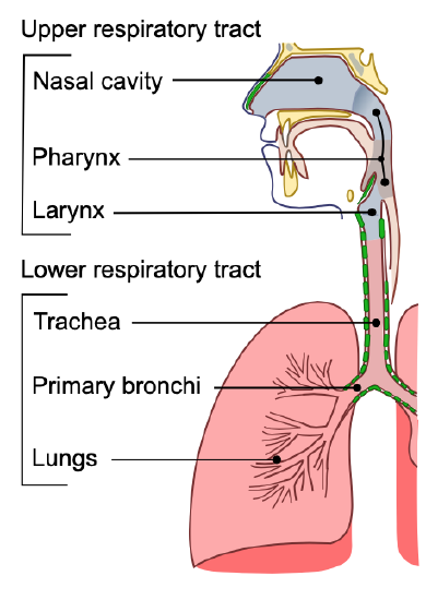
The organs of the respiratory system form a continuous system of passages called the respiratory tract, through which air flows into and out of the body. The respiratory tract has two major divisions: the upper respiratory tract and the lower respiratory tract. The organs in each division are shown in Figure \(\PageIndex{2}\). In addition to these organs, certain muscles of the thorax (the body cavity that fills the chest) are also involved in respiration by enabling breathing. Most important is a large muscle called the diaphragm, which lies below the lungs and separates the thorax from the abdomen. Smaller muscles between the ribs also play a role in breathing. You can learn more about breathing muscles in the concept of Breathing .
Upper Respiratory Tract
All of the organs and other structures of the upper respiratory tract are involved in the conduction or the movement of air into and out of the body. Upper respiratory tract organs provide a route for air to move between the outside atmosphere and the lungs. They also clean, humidity, and warm the incoming air. However, no gas exchange occurs in these organs.
Nasal Cavity
The nasal cavity is a large, air-filled space in the skull above and behind the nose in the middle of the face. It is a continuation of the two nostrils. As inhaled air flows through the nasal cavity, it is warmed and humidified. Hairs in the nose help trap larger foreign particles in the air before they go deeper into the respiratory tract. In addition to its respiratory functions, the nasal cavity also contains chemoreceptors that are needed for the sense of smell and that contribute importantly to the sense of taste.
The pharynx is a tube-like structure that connects the nasal cavity and the back of the mouth to other structures lower in the throat, including the larynx. The pharynx has dual functions: both air and food (or other swallowed substances) pass through it, so it is part of both the respiratory and digestive systems. Air passes from the nasal cavity through the pharynx to the larynx (as well as in the opposite direction). Food passes from the mouth through the pharynx to the esophagus.
The larynx connects the pharynx and trachea and helps to conduct air through the respiratory tract. The larynx is also called the voice box because it contains the vocal cords, which vibrate when air flows over them, thereby producing sound. You can see the vocal cords in the larynx in Figure \(\PageIndex{3}\). Certain muscles in the larynx move the vocal cords apart to allow breathing. Other muscles in the larynx move the vocal cords together to allow the production of vocal sounds. The latter muscles also control the pitch of sounds and help control their volume.
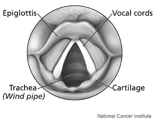
A very important function of the larynx is protecting the trachea from aspirated food. When swallowing occurs, the backward motion of the tongue forces a flap called the epiglottis to close over the entrance to the larynx. You can see the epiglottis in Figure \(\PageIndex{3}\). This prevents swallowed material from entering the larynx and moving deeper into the respiratory tract. If swallowed material does start to enter the larynx, it irritates the larynx and stimulates a strong cough reflex. This generally expels the material out of the larynx and into the throat.
Lower Respiratory Tract
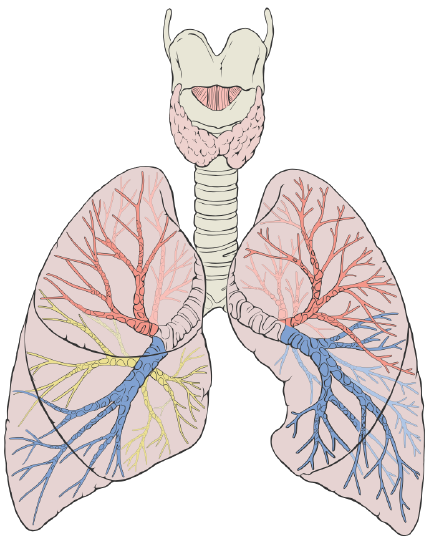
The trachea and other passages of the lower respiratory tract conduct air between the upper respiratory tract and the lungs. These passages form an inverted tree-like shape (Figure \(\PageIndex{4}\)), with repeated branching as they move deeper into the lungs. All told, there are an astonishing 1,500 miles of airways conducting air through the human respiratory tract! It is only in the lungs, however, that gas exchange occurs between the air and the bloodstream.
The trachea, or windpipe, is the widest passageway in the respiratory tract. It is about 2.5 cm (1 in.) wide and 10-15 cm (4-6 in.) long. It is formed by rings of cartilage, which make it relatively strong and resilient. The trachea connects the larynx to the lungs for the passage of air through the respiratory tract. The trachea branches at the bottom to form two bronchial tubes.
Bronchi and Bronchioles
There are two main bronchial tubes, or bronchi (singular, bronchus) , called the right and left bronchi. The bronchi carry air between the trachea and lungs. Each bronchus branches into smaller, secondary bronchi; and secondary bronchi branch into still smaller tertiary bronchi. The smallest bronchi branch into very small tubules called bronchioles. The tiniest bronchioles end in alveolar ducts, which terminate in clusters of minuscule air sacs, called alveoli (singular, alveolus), in the lungs.
The lungs are the largest organs of the respiratory tract. They are suspended within the pleural cavity of the thorax. In Figure \(\PageIndex{5}\), you can see that each of the two lungs is divided into sections. These are called lobes, and they are separated from each other by connective tissues. The right lung is larger and contains three lobes. The left lung is smaller and contains only two lobes. The smaller left lung allows room for the heart, which is just left of the center of the chest.
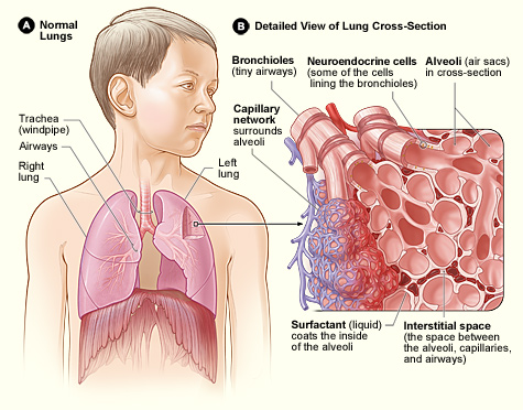
Lung tissue consists mainly of alveoli (Figure \(\PageIndex{6}\)). These tiny air sacs are the functional units of the lungs where gas exchange takes place. The two lungs may contain as many as 700 million alveoli, providing a huge total surface area for gas exchange to take place. In fact, alveoli in the two lungs provide as much surface area as half a tennis court! Each time you breathe in, the alveoli fill with air, making the lungs expand. Oxygen in the air inside the alveoli is absorbed by the blood in the mesh-like network of tiny capillaries that surrounds each alveolus. The blood in these capillaries also releases carbon dioxide into the air inside the alveoli. Each time you breathe out, air leaves the alveoli and rushes into the outside atmosphere, carrying waste gases with it.
The lungs receive blood from two major sources. They receive deoxygenated blood from the heart. This blood absorbs oxygen in the lungs and carries it back to the heart to be pumped to cells throughout the body. The lungs also receive oxygenated blood from the heart that provides oxygen to the cells of the lungs for cellular respiration.
Protecting the Respiratory System
You may be able to survive for weeks without food and for days without water, but you can survive without oxygen for only a matter of minutes except under exceptional circumstances. Therefore, protecting the respiratory system is vital. That’s why making sure a patient has an open airway is the first step in treating many medical emergencies. Fortunately, the respiratory system is well protected by the ribcage of the skeletal system. However, the extensive surface area of the respiratory system is directly exposed to the outside world and all its potential dangers in inhaled air. Therefore, it should come as no surprise that the respiratory system has a variety of ways to protect itself from harmful substances such as dust and pathogens in the air.
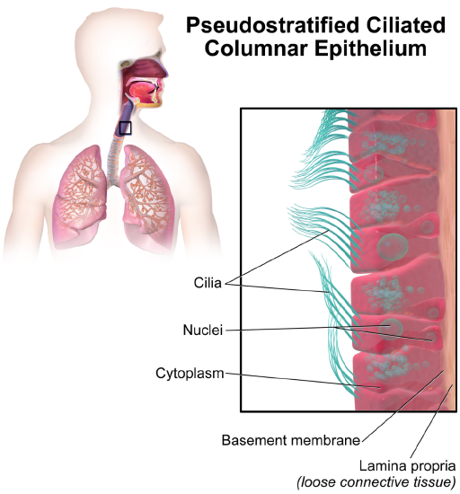
The main way the respiratory system protects itself is called the mucociliary escalator. From the nose through the bronchi, the respiratory tract is covered in the epithelium that contains mucus-secreting goblet cells. The mucus traps particles and pathogens in the incoming air. The epithelium of the respiratory tract is also covered with tiny cell projections called cilia (singular, cilium), as shown in Figure \(\PageIndex{7}\). The cilia constantly move in a sweeping motion upward toward the throat, moving the mucus and trapped particles and pathogens away from the lungs and toward the outside of the body.
What happens to the material that moves up the mucociliary escalator to the throat? It is generally removed from the respiratory tract by clearing the throat or coughing. Coughing is a largely involuntary response of the respiratory system that occurs when nerves lining the airways are irritated. The response causes air to be expelled forcefully from the trachea, helping to remove mucus and any debris it contains (called phlegm) from the upper respiratory tract to the mouth. The phlegm may spit out (expectorated), or it may be swallowed and destroyed by stomach acids.
Sneezing is a similar involuntary response that occurs when nerves lining the nasal passage are irritated. It results in forceful expulsion of air from the mouth, which sprays millions of tiny droplets of mucus and other debris out of the mouth and into the air, as shown in Figure \(\PageIndex{8}\). This explains why it is so important to sneeze into a sleeve rather than the air to help prevent the transmission of respiratory pathogens.
How the Respiratory System Works with Other Organ Systems
The amount of oxygen and carbon dioxide in the blood must be maintained within a limited range for the survival of the organism. Cells cannot survive for long without oxygen, and if there is too much carbon dioxide in the blood, the blood becomes dangerously acidic (pH is too low). Conversely, if there is too little carbon dioxide in the blood, the blood becomes too basic (pH is too high). The respiratory system works hand-in-hand with the nervous and cardiovascular systems to maintain homeostasis in blood gases and pH.
It is the level of carbon dioxide rather than the level of oxygen that is most closely monitored to maintain blood gas and pH homeostasis. The level of carbon dioxide in the blood is detected by cells in the brain, which speed up or slow down the rate of breathing through the autonomic nervous system as needed to bring the carbon dioxide level within the normal range. Faster breathing lowers the carbon dioxide level (and raises the oxygen level and pH); slower breathing has the opposite effects. In this way, the levels of carbon dioxide and oxygen, as well as pH, are maintained within normal limits.
The respiratory system also works closely with the cardiovascular system to maintain homeostasis. The respiratory system exchanges gases between the blood and the outside air, but it needs the cardiovascular system to carry them to and from body cells. Oxygen is absorbed by the blood in the lungs and then transported through a vast network of blood vessels to cells throughout the body where it is needed for aerobic cellular respiration. The same system absorbs carbon dioxide from cells and carries it to the respiratory system for removal from the body.
Feature: My Human Body
Choking is the mechanical obstruction of the flow of air from the atmosphere into the lungs. It prevents breathing and may be partial or complete. Partial choking allows some though inadequate airflow into the lung—prolonged or complete choking results in asphyxia, or suffocation, which is potentially fatal.
Obstruction of the airway typically occurs in the pharynx or trachea. Young children are more prone to choking than are older people, in part because they often put small objects in their mouths and do not appreciate the risk of choking that they pose. Young children may choke on small toys or parts of toys or on household objects in addition to food. Foods that can adapt their shape to that of the pharynx, such as bananas and marshmallows, are especially dangerous and may cause choking in adults as well as children.
How can you tell if a loved one is choking? The person cannot speak or cry out or has great difficulty doing so. Breathing, if possible, is labored, producing gasping or wheezing. The person may desperately clutch at his or her throat or mouth. If breathing is not soon restored, the person’s face will start to turn blue from lack of oxygen. This will be followed by unconsciousness if oxygen deprivation continues beyond a few minutes.
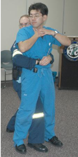
If an infant is choking, turning the baby upside down and slapping on the back may dislodge the obstructing object. To help an older person who is choking, first, encourage the person to cough. Give them a few hardback slaps to help force the lodged object out of the airway. If these steps fail, perform the Heimlich maneuver on the person. You can easily find instructional videos online to learn how to do it. If the Heimlich maneuver also fails, call for emergency medical care immediately.
- What is respiration, as carried out by the respiratory system? Name the two subsidiary processes it involves.
- Describe the respiratory tract.
- Identify the organs of the upper respiratory tract, and state their functions.
- List the organs of the lower respiratory tract. Which organs are involved only in conduction?
- Where does gas exchange take place?
- How does the respiratory system protect itself from potentially harmful substances in the air?
- Explain how the rate of breathing is controlled.
- Why does the respiratory system need the cardiovascular system to help it perform its main function of gas exchange?
trachea; nasal cavity; alveoli; bronchioles; larynx; bronchi; pharynx
D. Bronchus
- Describe two ways in which the body prevents food from entering the lungs.
- True or False. The lungs receive some oxygenated blood.
- True or False. Gas exchange occurs in both the upper and lower respiratory tracts.
B. food particles
D. All of the above
- What is the relationship between respiration and cellular respiration?
Explore More
Attributions.
- Snowboarders breath on a cold day by Alain Wong via Unsplash License
- Conducting Passages by Lord Akryl , Jmarchn, public domain via Wikimedia Commons
- Larynx by Alan Hoofring , National Cancer Institute, public domain via Wikimedia Commons
- Lung Diagram by Patrick J. Lynch ; CC BY 2.5 via Wikimedia Commons
- Lung Structure by National Heart Lung and Blood Institute, public domain via Wikimedia Commons
- Alveoli by helix84 licensed CC BY 2.5 , via Wikimedia Commons
- Ciliated Epithelium by Blausen.com staff (2014). " Medical gallery of Blausen Medical 2014 ". WikiJournal of Medicine 1 (2). DOI : 10.15347/wjm/2014.010 . ISSN 2002-4436 . licensed CC BY 3.0 via Wikimedia Commons
- Sneeze by James Gathany, CDC , public domain via Wikimedia Commons
- Abdominal Thrusts by Amanda M. Woodhead, public domain via Wikimedia Commons
- Text adapted from Human Biology by CK-12 licensed CC BY-NC 3.0
16.3 Circulatory and Respiratory Systems
Learning objectives.
- Describe the passage of air from the outside environment to the lungs
- Describe the function of the circulatory system
- Describe the cardiac cycle
- Explain how blood flows through the body
Animals are complex multicellular organisms that require a mechanism for transporting nutrients throughout their bodies and removing wastes. The human circulatory system has a complex network of blood vessels that reach all parts of the body. This extensive network supplies the cells, tissues, and organs with oxygen and nutrients, and removes carbon dioxide and waste compounds.
The medium for transport of gases and other molecules is the blood, which continually circulates through the system. Pressure differences within the system cause the movement of the blood and are created by the pumping of the heart.
Gas exchange between tissues and the blood is an essential function of the circulatory system. In humans, other mammals, and birds, blood absorbs oxygen and releases carbon dioxide in the lungs. Thus the circulatory and respiratory system, whose function is to obtain oxygen and discharge carbon dioxide, work in tandem.
The Respiratory System
Take a breath in and hold it. Wait several seconds and then let it out. Humans, when they are not exerting themselves, breathe approximately 15 times per minute on average. This equates to about 900 breaths an hour or 21,600 breaths per day. With every inhalation, air fills the lungs, and with every exhalation, it rushes back out. That air is doing more than just inflating and deflating the lungs in the chest cavity. The air contains oxygen that crosses the lung tissue, enters the bloodstream, and travels to organs and tissues. There, oxygen is exchanged for carbon dioxide, which is a cellular waste material. Carbon dioxide exits the cells, enters the bloodstream, travels back to the lungs, and is expired out of the body during exhalation.
Breathing is both a voluntary and an involuntary event. How often a breath is taken and how much air is inhaled or exhaled is regulated by the respiratory center in the brain in response to signals it receives about the carbon dioxide content of the blood. However, it is possible to override this automatic regulation for activities such as speaking, singing and swimming under water.
During inhalation the diaphragm descends creating a negative pressure around the lungs and they begin to inflate, drawing in air from outside the body. The air enters the body through the nasal cavity located just inside the nose ( Figure 16.9 ). As the air passes through the nasal cavity, the air is warmed to body temperature and humidified by moisture from mucous membranes. These processes help equilibrate the air to the body conditions, reducing any damage that cold, dry air can cause. Particulate matter that is floating in the air is removed in the nasal passages by hairs, mucus, and cilia. Air is also chemically sampled by the sense of smell.
From the nasal cavity, air passes through the pharynx (throat) and the larynx (voice box) as it makes its way to the trachea ( Figure 16.9 ). The main function of the trachea is to funnel the inhaled air to the lungs and the exhaled air back out of the body. The human trachea is a cylinder, about 25 to 30 cm (9.8–11.8 in) long, which sits in front of the esophagus and extends from the pharynx into the chest cavity to the lungs. It is made of incomplete rings of cartilage and smooth muscle. The cartilage provides strength and support to the trachea to keep the passage open. The trachea is lined with cells that have cilia and secrete mucus. The mucus catches particles that have been inhaled, and the cilia move the particles toward the pharynx.
The end of the trachea divides into two bronchi that enter the right and left lung. Air enters the lungs through the primary bronchi . The primary bronchus divides, creating smaller and smaller diameter bronchi until the passages are under 1 mm (.03 in) in diameter when they are called bronchioles as they split and spread through the lung. Like the trachea, the bronchus and bronchioles are made of cartilage and smooth muscle. Bronchi are innervated by nerves of both the parasympathetic and sympathetic nervous systems that control muscle contraction (parasympathetic) or relaxation (sympathetic) in the bronchi and bronchioles, depending on the nervous system’s cues. The final bronchioles are the respiratory bronchioles. Alveolar ducts are attached to the end of each respiratory bronchiole. At the end of each duct are alveolar sacs, each containing 20 to 30 alveoli . Gas exchange occurs only in the alveoli. The alveoli are thin-walled and look like tiny bubbles within the sacs. The alveoli are in direct contact with capillaries of the circulatory system. Such intimate contact ensures that oxygen will diffuse from the alveoli into the blood. In addition, carbon dioxide will diffuse from the blood into the alveoli to be exhaled. The anatomical arrangement of capillaries and alveoli emphasizes the structural and functional relationship of the respiratory and circulatory systems. Estimates for the surface area of alveoli in the lungs vary around 100 m 2 . This large area is about the area of half a tennis court. This large surface area, combined with the thin-walled nature of the alveolar cells, allows gases to easily diffuse across the cells.
Visual Connection
Which of the following statements about the human respiratory system is false?
- When we breathe in, air travels from the pharynx to the trachea.
- The bronchioles branch into bronchi.
- Alveolar ducts connect to alveolar sacs.
- Gas exchange between the lungs and blood takes place in the alveolus.
Link to Learning
Watch this video for a review of the respiratory system.
The Circulatory System
The circulatory system is a network of vessels—the arteries, veins, and capillaries—and a pump, the heart. In all vertebrate organisms this is a closed-loop system, in which the blood is largely separated from the body’s other extracellular fluid compartment, the interstitial fluid, which is the fluid bathing the cells. Blood circulates inside blood vessels and circulates unidirectionally from the heart around one of two circulatory routes, then returns to the heart again; this is a closed circulatory system . Open circulatory systems are found in invertebrate animals in which the circulatory fluid bathes the internal organs directly even though it may be moved about with a pumping heart.
The heart is a complex muscle that consists of two pumps: one that pumps blood through pulmonary circulation to the lungs, and the other that pumps blood through systemic circulation to the rest of the body’s tissues (and the heart itself).
The heart is asymmetrical, with the left side being larger than the right side, correlating with the different sizes of the pulmonary and systemic circuits ( Figure 16.10 ). In humans, the heart is about the size of a clenched fist; it is divided into four chambers: two atria and two ventricles. There is one atrium and one ventricle on the right side and one atrium and one ventricle on the left side. The right atrium receives deoxygenated blood from the systemic circulation through the major veins: the superior vena cava , which drains blood from the head and from the veins that come from the arms, as well as the inferior vena cava , which drains blood from the veins that come from the lower organs and the legs. This deoxygenated blood then passes to the right ventricle through the tricuspid valve , which prevents the backflow of blood. After it is filled, the right ventricle contracts, pumping the blood to the lungs for reoxygenation. The left atrium receives the oxygen-rich blood from the lungs. This blood passes through the bicuspid valve to the left ventricle where the blood is pumped into the aorta . The aorta is the major artery of the body, taking oxygenated blood to the organs and muscles of the body. This pattern of pumping is referred to as double circulation and is found in all mammals. ( Figure 16.10 ).
Which of the following statements about the circulatory system is false?
- Blood in the pulmonary vein is deoxygenated.
- Blood in the inferior vena cava is deoxygenated.
- Blood in the pulmonary artery is deoxygenated.
- Blood in the aorta is oxygenated.
The Cardiac Cycle
The main purpose of the heart is to pump blood through the body; it does so in a repeating sequence called the cardiac cycle. The cardiac cycle is the flow of blood through the heart coordinated by electrochemical signals that cause the heart muscle to contract and relax. In each cardiac cycle, a sequence of contractions pushes out the blood, pumping it through the body; this is followed by a relaxation phase, where the heart fills with blood. These two phases are called the systole (contraction) and diastole (relaxation), respectively ( Figure 16.11 ). The signal for contraction begins at a location on the outside of the right atrium. The electrochemical signal moves from there across the atria causing them to contract. The contraction of the atria forces blood through the valves into the ventricles. Closing of these valves caused by the contraction of the ventricles produces a “lub” sound. The signal has, by this time, passed down the walls of the heart, through a point between the right atrium and right ventricle. The signal then causes the ventricles to contract. The ventricles contract together forcing blood into the aorta and the pulmonary arteries. Closing of the valves to these arteries caused by blood being drawn back toward the heart during ventricular relaxation produces a monosyllabic “dub” sound.
The pumping of the heart is a function of the cardiac muscle cells, or cardiomyocytes, that make up the heart muscle. Cardiomyocytes are distinctive muscle cells that are striated like skeletal muscle but pump rhythmically and involuntarily like smooth muscle; adjacent cells are connected by intercalated disks found only in cardiac muscle. These connections allow the electrical signal to travel directly to neighboring muscle cells.
The electrical impulses in the heart produce electrical currents that flow through the body and can be measured on the skin using electrodes. This information can be observed as an electrocardiogram (ECG) a recording of the electrical impulses of the cardiac muscle.
Visit this site and select the dropdown “Your Heart’s Electrical System” to see the heart’s pacemaker, or electrocardiogram system, in action.
Blood Vessels
The blood from the heart is carried through the body by a complex network of blood vessels ( Figure 16.12 ). Arteries take blood away from the heart. The main artery of the systemic circulation is the aorta; it branches into major arteries that take blood to different limbs and organs. The aorta and arteries near the heart have heavy but elastic walls that respond to and smooth out the pressure differences caused by the beating heart. Arteries farther away from the heart have more muscle tissue in their walls that can constrict to affect flow rates of blood. The major arteries diverge into minor arteries, and then smaller vessels called arterioles, to reach more deeply into the muscles and organs of the body.
Arterioles diverge into capillary beds. Capillary beds contain a large number, 10’s to 100’s of capillaries that branch among the cells of the body. Capillaries are narrow-diameter tubes that can fit single red blood cells and are the sites for the exchange of nutrients, waste, and oxygen with tissues at the cellular level. Fluid also leaks from the blood into the interstitial space from the capillaries. The capillaries converge again into venules that connect to minor veins that finally connect to major veins. Veins are blood vessels that bring blood high in carbon dioxide back to the heart. Veins are not as thick-walled as arteries, since pressure is lower, and they have valves along their length that prevent backflow of blood away from the heart. The major veins drain blood from the same organs and limbs that the major arteries supply.
As an Amazon Associate we earn from qualifying purchases.
This book may not be used in the training of large language models or otherwise be ingested into large language models or generative AI offerings without OpenStax's permission.
Want to cite, share, or modify this book? This book uses the Creative Commons Attribution License and you must attribute OpenStax.
Access for free at https://openstax.org/books/concepts-biology/pages/1-introduction
- Authors: Samantha Fowler, Rebecca Roush, James Wise
- Publisher/website: OpenStax
- Book title: Concepts of Biology
- Publication date: Apr 25, 2013
- Location: Houston, Texas
- Book URL: https://openstax.org/books/concepts-biology/pages/1-introduction
- Section URL: https://openstax.org/books/concepts-biology/pages/16-3-circulatory-and-respiratory-systems
© Jan 8, 2024 OpenStax. Textbook content produced by OpenStax is licensed under a Creative Commons Attribution License . The OpenStax name, OpenStax logo, OpenStax book covers, OpenStax CNX name, and OpenStax CNX logo are not subject to the Creative Commons license and may not be reproduced without the prior and express written consent of Rice University.

- school Campus Bookshelves
- menu_book Bookshelves
- perm_media Learning Objects
- login Login
- how_to_reg Request Instructor Account
- hub Instructor Commons
- Download Page (PDF)
- Download Full Book (PDF)
- Periodic Table
- Physics Constants
- Scientific Calculator
- Reference & Cite
- Tools expand_more
- Readability
selected template will load here
This action is not available.

14.2: Organs and Structures of the Respiratory System
- Last updated
- Save as PDF
- Page ID 57551
Learning Objectives
- List the structures that make up the respiratory system
- Describe how the respiratory system processes oxygen and CO 2
- Compare and contrast the functions of upper respiratory tract with the lower respiratory tract
The major organs of the respiratory system function primarily to provide oxygen to body tissues for cellular respiration, remove the waste product carbon dioxide, and help to maintain acid-base balance. Portions of the respiratory system are also used for non-vital functions, such as sensing odors, speech production, and for straining, such as during childbirth or coughing (Figure \(\PageIndex{1}\)).

Functionally, the respiratory system can be divided into a conducting zone and a respiratory zone. The conducting zone of the respiratory system includes the organs and structures not directly involved in gas exchange. The gas exchange occurs in the respiratory zone .
Conducting Zone
The major functions of the conducting zone are to provide a route for incoming and outgoing air, remove debris and pathogens from the incoming air, and warm and humidify the incoming air. Several structures within the conducting zone perform other functions as well. The epithelium of the nasal passages, for example, is essential to sensing odors, and the bronchial epithelium that lines the lungs can metabolize some airborne carcinogens.
The Nose and Its Adjacent Structures
The major entrance and exit for the respiratory system is through the nose. When discussing the nose, it is helpful to divide it into two major sections: the external nose, and the nasal cavity or internal nose.
The external nose consists of the surface and skeletal structures that result in the outward appearance of the nose and contribute to its numerous functions (Figure \(\PageIndex{2}\)). The root is the region of the nose located between the eyebrows. The bridge is the part of the nose that connects the root to the rest of the nose. The dorsum nasi is the length of the nose. The apex is the tip of the nose. On either side of the apex, the nostrils are formed by the alae (singular = ala). An ala is a cartilaginous structure that forms the lateral side of each naris (plural = nares), or nostril opening. The philtrum is the concave surface that connects the apex of the nose to the upper lip.

Underneath the thin skin of the nose are its skeletal features (see Figure \(\PageIndex{2}\), lower illustration). While the root and bridge of the nose consist of bone, the protruding portion of the nose is composed of cartilage. As a result, when looking at a skull, the nose is missing. The nasal bone is one of a pair of bones that lies under the root and bridge of the nose. The nasal bone articulates superiorly with the frontal bone and laterally with the maxillary bones. Septal cartilage is flexible hyaline cartilage connected to the nasal bone, forming the dorsum nasi. The alar cartilage consists of the apex of the nose; it surrounds the naris.
The nares open into the nasal cavity, which is separated into left and right sections by the nasal septum (Figure \(\PageIndex{3}\)). The nasal septum is formed anteriorly by a portion of the septal cartilage (the flexible portion you can touch with your fingers) and posteriorly by the perpendicular plate of the ethmoid bone (a cranial bone located just posterior to the nasal bones) and the thin vomer bones (whose name refers to its plough shape). Each lateral wall of the nasal cavity has three bony projections, called the superior, middle, and inferior nasal conchae. The inferior conchae are separate bones, whereas the superior and middle conchae are portions of the ethmoid bone. Conchae serve to increase the surface area of the nasal cavity and to disrupt the flow of air as it enters the nose, causing air to bounce along the epithelium, where it is cleaned and warmed. The conchae and meatuses also conserve water and prevent dehydration of the nasal epithelium by trapping water during exhalation. The floor of the nasal cavity is composed of the palate. The hard palate at the anterior region of the nasal cavity is composed of bone. The soft palate at the posterior portion of the nasal cavity consists of muscle tissue. Air exits the nasal cavities via the internal nares and moves into the pharynx.

Several bones that help form the walls of the nasal cavity have air-containing spaces called the paranasal sinuses, which serve to warm and humidify incoming air. Sinuses are lined with a mucosa. Each paranasal sinus is named for its associated bone: frontal sinus, maxillary sinus, sphenoidal sinus, and ethmoidal sinus. The sinuses produce mucus and lighten the weight of the skull.
The nares and anterior portion of the nasal cavities are lined with mucous membranes, containing sebaceous glands and hair follicles that serve to prevent the passage of large debris, such as dirt, through the nasal cavity. An olfactory epithelium used to detect odors is found deeper in the nasal cavity.
The conchae, meatuses, and paranasal sinuses are lined by respiratory epithelium composed of pseudostratified ciliated columnar epithelium (Figure \(\PageIndex{4}\)). The epithelium contains goblet cells, one of the specialized, columnar epithelial cells that produce mucus to trap debris. The cilia of the respiratory epithelium help remove the mucus and debris from the nasal cavity with a constant beating motion, sweeping materials towards the throat to be swallowed. Interestingly, cold air slows the movement of the cilia, resulting in accumulation of mucus that may in turn lead to a runny nose during cold weather. This moist epithelium functions to warm and humidify incoming air. Capillaries located just beneath the nasal epithelium warm the air by convection. Serous and mucus-producing cells also secrete the lysozyme enzyme and proteins called defensins, which have antibacterial properties. Immune cells that patrol the connective tissue deep to the respiratory epithelium provide additional protection.

The pharynx is a tube formed by skeletal muscle and lined by mucous membrane that is continuous with that of the nasal cavities (see Figure \(\PageIndex{3}\)). The pharynx is divided into three major regions: the nasopharynx, the oropharynx, and the laryngopharynx (Figure \(\PageIndex{5}\)).

The nasopharynx is flanked by the conchae of the nasal cavity, and it serves only as an airway. At the top of the nasopharynx are the pharyngeal tonsils. A pharyngeal tonsil , also called an adenoid, is an aggregate of lymphoid reticular tissue similar to a lymph node that lies at the superior portion of the nasopharynx. The function of the pharyngeal tonsil is not well understood, but it contains a rich supply of lymphocytes and is covered with ciliated epithelium that traps and destroys invading pathogens that enter during inhalation. The pharyngeal tonsils are large in children, but interestingly, tend to regress with age and may even disappear. The uvula is a small bulbous, teardrop-shaped structure located at the apex of the soft palate. Both the uvula and soft palate move like a pendulum during swallowing, swinging upward to close off the nasopharynx to prevent ingested materials from entering the nasal cavity. In addition, auditory (Eustachian) tubes that connect to each middle ear cavity open into the nasopharynx. This connection is why colds often lead to ear infections.
The oropharynx is a passageway for both air and food. The oropharynx is bordered superiorly by the nasopharynx and anteriorly by the oral cavity. The fauces is the opening at the connection between the oral cavity and the oropharynx. As the nasopharynx becomes the oropharynx, the epithelium changes from pseudostratified ciliated columnar epithelium to stratified squamous epithelium. The oropharynx contains two distinct sets of tonsils, the palatine and lingual tonsils. A palatine tonsil is one of a pair of structures located laterally in the oropharynx in the area of the fauces. The lingual tonsil is located at the base of the tongue. Similar to the pharyngeal tonsil, the palatine and lingual tonsils are composed of lymphoid tissue, and trap and destroy pathogens entering the body through the oral or nasal cavities.
The laryngopharynx is inferior to the oropharynx and posterior to the larynx. It continues the route for ingested material and air until its inferior end, where the digestive and respiratory systems diverge. The stratified squamous epithelium of the oropharynx is continuous with the laryngopharynx. Anteriorly, the laryngopharynx opens into the larynx, whereas posteriorly, it enters the esophagus.
The larynx is a cartilaginous structure inferior to the laryngopharynx that connects the pharynx to the trachea and helps regulate the volume of air that enters and leaves the lungs (Figure \(\PageIndex{6}\)). The structure of the larynx is formed by several pieces of cartilage. Three large cartilage pieces—the thyroid cartilage (anterior), epiglottis (superior), and cricoid cartilage (inferior)—form the major structure of the larynx. The thyroid cartilage is the largest piece of cartilage that makes up the larynx. The thyroid cartilage consists of the laryngeal prominence , or “Adam’s apple,” which tends to be more prominent in males. The thick cricoid cartilage forms a ring, with a wide posterior region and a thinner anterior region. Three smaller, paired cartilages—the arytenoids, corniculates, and cuneiforms—attach to the epiglottis and the vocal cords and muscle that help move the vocal cords to produce speech.

The epiglottis , attached to the thyroid cartilage, is a very flexible piece of elastic cartilage that covers the opening of the trachea (see Figure \(\PageIndex{3}\)). When in the “closed” position, the unattached end of the epiglottis rests on the glottis. The glottis is composed of the vestibular folds, the true vocal cords, and the space between these folds (Figure \(\PageIndex{7}\)). A vestibular fold , or false vocal cord, is one of a pair of folded sections of mucous membrane. A true vocal cord is one of the white, membranous folds attached by muscle to the thyroid and arytenoid cartilages of the larynx on their outer edges. The inner edges of the true vocal cords are free, allowing oscillation to produce sound. The size of the membranous folds of the true vocal cords differs between individuals, producing voices with different pitch ranges. Folds in males tend to be larger than those in females, which create a deeper voice. The act of swallowing causes the pharynx and larynx to lift upward, allowing the pharynx to expand and the epiglottis of the larynx to swing downward, closing the opening to the trachea. These movements produce a larger area for food to pass through, while preventing food and beverages from entering the trachea.

Continuous with the laryngopharynx, the superior portion of the larynx is lined with stratified squamous epithelium, transitioning into pseudostratified ciliated columnar epithelium that contains goblet cells. Similar to the nasal cavity and nasopharynx, this specialized epithelium produces mucus to trap debris and pathogens as they enter the trachea. The cilia beat the mucus upward towards the laryngopharynx, where it can be swallowed down the esophagus.
The trachea (windpipe) extends from the larynx toward the lungs (Figure \(\PageIndex{8}\).a). The trachea is formed by 16 to 20 stacked, C-shaped pieces of hyaline cartilage that are connected by dense connective tissue. The trachealis muscle and elastic connective tissue together form the fibroelastic membrane , a flexible membrane that closes the posterior surface of the trachea, connecting the C-shaped cartilages. The fibroelastic membrane allows the trachea to stretch and expand slightly during inhalation and exhalation, whereas the rings of cartilage provide structural support and prevent the trachea from collapsing. In addition, the trachealis muscle can be contracted to force air through the trachea during exhalation. The trachea is lined with pseudostratified ciliated columnar epithelium, which is continuous with the larynx. The esophagus borders the trachea posteriorly.


Bronchial Tree
The trachea branches into the right and left primary bronchi at the carina. These bronchi are also lined by pseudostratified ciliated columnar epithelium containing mucus-producing goblet cells (Figure \(\PageIndex{8}\).b). The carina is a raised structure that contains specialized nervous tissue that induces violent coughing if a foreign body, such as food, is present. Rings of cartilage, similar to those of the trachea, support the structure of the bronchi and prevent their collapse. The primary bronchi enter the lungs at the hilum, a concave region where blood vessels, lymphatic vessels, and nerves also enter the lungs. The bronchi continue to branch into bronchial a tree. A bronchial tree (or respiratory tree) is the collective term used for these multiple-branched bronchi. The main function of the bronchi, like other conducting zone structures, is to provide a passageway for air to move into and out of each lung. In addition, the mucous membrane traps debris and pathogens.
A bronchiole branches from the tertiary bronchi. Bronchioles, which are about 1 mm in diameter, further branch until they become the tiny terminal bronchioles, which lead to the structures of gas exchange. There are more than 1000 terminal bronchioles in each lung. The muscular walls of the bronchioles do not contain cartilage like those of the bronchi. This muscular wall can change the size of the tubing to increase or decrease airflow through the tube.
Respiratory Zone
In contrast to the conducting zone, the respiratory zone includes structures that are directly involved in gas exchange. The respiratory zone begins where the terminal bronchioles join a respiratory bronchiole , the smallest type of bronchiole (Figure \(\PageIndex{9}\)), which then leads to an alveolar duct, opening into a cluster of alveoli.

An alveolar duct is a tube composed of smooth muscle and connective tissue, which opens into a cluster of alveoli. An alveolus is one of the many small, grape-like sacs that are attached to the alveolar ducts.
An alveolar sac is a cluster of many individual alveoli that are responsible for gas exchange. An alveolus is approximately 200 μm in diameter with elastic walls that allow the alveolus to stretch during air intake, which greatly increases the surface area available for gas exchange. Alveoli are connected to their neighbors by alveolar pores , which help maintain equal air pressure throughout the alveoli and lung (Figure \(\PageIndex{10}\)).

The alveolar wall consists of three major cell types: type I alveolar cells, type II alveolar cells, and alveolar macrophages. A type I alveolar cell is a squamous epithelial cell of the alveoli, which constitute up to 97 percent of the alveolar surface area. These cells are about 25 nm thick and are highly permeable to gases. A type II alveolar cell is interspersed among the type I cells and secretes pulmonary surfactant , a substance composed of phospholipids and proteins that reduces the surface tension of the alveoli. Roaming around the alveolar wall is the alveolar macrophage , a phagocytic cell of the immune system that removes debris and pathogens that have reached the alveoli.
The simple squamous epithelium formed by type I alveolar cells is attached to a thin, elastic basement membrane. This epithelium is extremely thin and borders the endothelial membrane of capillaries. Taken together, the alveoli and capillary membranes form a respiratory membrane that is approximately 0.5 mm thick. The respiratory membrane allows gases to cross by simple diffusion, allowing oxygen to be picked up by the blood for transport and CO 2 to be released into the air of the alveoli.
Diseases of the Respiratory System: Asthma
Asthma is common condition that affects the lungs in both adults and children. Approximately 8.2 percent of adults (18.7 million) and 9.4 percent of children (7 million) in the United States suffer from asthma. In addition, asthma is the most frequent cause of hospitalization in children.
Asthma is a chronic disease characterized by inflammation and edema of the airway, and bronchospasms (that is, constriction of the bronchioles), which can inhibit air from entering the lungs. In addition, excessive mucus secretion can occur, which further contributes to airway occlusion (Figure \(\PageIndex{11}\)). Cells of the immune system, such as eosinophils and mononuclear cells, may also be involved in infiltrating the walls of the bronchi and bronchioles.
Bronchospasms occur periodically and lead to an “asthma attack.” An attack may be triggered by environmental factors such as dust, pollen, pet hair, or dander, changes in the weather, mold, tobacco smoke, and respiratory infections, or by exercise and stress.

Symptoms of an asthma attack involve coughing, shortness of breath, wheezing, and tightness of the chest. Symptoms of a severe asthma attack that requires immediate medical attention would include difficulty breathing that results in blue (cyanotic) lips or face, confusion, drowsiness, a rapid pulse, sweating, and severe anxiety. The severity of the condition, frequency of attacks, and identified triggers influence the type of medication that an individual may require. Longer-term treatments are used for those with more severe asthma. Short-term, fast-acting drugs that are used to treat an asthma attack are typically administered via an inhaler. For young children or individuals who have difficulty using an inhaler, asthma medications can be administered via a nebulizer.
In many cases, the underlying cause of the condition is unknown. However, recent research has demonstrated that certain viruses, such as human rhinovirus C (HRVC), and the bacteria Mycoplasma pneumoniae and Chlamydia pneumoniae that are contracted in infancy or early childhood, may contribute to the development of many cases of asthma.
Chapter Review
The respiratory system is responsible for obtaining oxygen and getting rid of carbon dioxide, and aiding in speech production and in sensing odors. From a functional perspective, the respiratory system can be divided into two major areas: the conducting zone and the respiratory zone. The conducting zone consists of all of the structures that provide passageways for air to travel into and out of the lungs: the nasal cavity, pharynx, trachea, bronchi, and most bronchioles. The nasal passages contain the conchae and meatuses that expand the surface area of the cavity, which helps to warm and humidify incoming air, while removing debris and pathogens. The pharynx is composed of three major sections: the nasopharynx, which is continuous with the nasal cavity; the oropharynx, which borders the nasopharynx and the oral cavity; and the laryngopharynx, which borders the oropharynx, trachea, and esophagus. The respiratory zone includes the structures of the lung that are directly involved in gas exchange: the terminal bronchioles and alveoli.
The lining of the conducting zone is composed mostly of pseudostratified ciliated columnar epithelium with goblet cells. The mucus traps pathogens and debris, whereas beating cilia move the mucus superiorly toward the throat, where it is swallowed. As the bronchioles become smaller and smaller, and nearer the alveoli, the epithelium thins and is simple squamous epithelium in the alveoli. The endothelium of the surrounding capillaries, together with the alveolar epithelium, forms the respiratory membrane. This is a blood-air barrier through which gas exchange occurs by simple diffusion.
Essay Questions
Q. What happens during an asthma attack. What are the three changes that occur inside the airways during an asthma attack?
A. Inflammation and the production of a thick mucus; constriction of the airway muscles, or bronchospasm; and an increased sensitivity to allergens.
Review Questions
Q. Which of the following anatomical structures is not part of the conducting zone?
B. nasal cavity
Q. What is the function of the conchae in the nasal cavity?
A. increase surface area
B. exchange gases
C. maintain surface tension
D. maintain air pressure
Q. The fauces connects which of the following structures to the oropharynx?
A. nasopharynx
B. laryngopharynx
C. nasal cavity
D. oral cavity
Q. Which of the following are structural features of the trachea?
A. C-shaped cartilage
B. smooth muscle fibers
D. all of the above
Q. Which of the following structures is not part of the bronchial tree?
C. terminal bronchioles
D. respiratory bronchioles
Q. What is the role of alveolar macrophages?
A. to secrete pulmonary surfactant
B. to secrete antimicrobial proteins
C. to remove pathogens and debris
D. to facilitate gas exchange
Critical Thinking Questions
Q. Describe the three regions of the pharynx and their functions.
A. The pharynx has three major regions. The first region is the nasopharynx, which is connected to the posterior nasal cavity and functions as an airway. The second region is the oropharynx, which is continuous with the nasopharynx and is connected to the oral cavity at the fauces. The laryngopharynx is connected to the oropharynx and the esophagus and trachea. Both the oropharynx and laryngopharynx are passageways for air and food and drink.
Q. If a person sustains an injury to the epiglottis, what would be the physiological result?
A. The epiglottis is a region of the larynx that is important during the swallowing of food or drink. As a person swallows, the pharynx moves upward and the epiglottis closes over the trachea, preventing food or drink from entering the trachea. If a person’s epiglottis were injured, this mechanism would be impaired. As a result, the person may have problems with food or drink entering the trachea, and possibly, the lungs. Over time, this may cause infections such as pneumonia to set in.
Q. Compare and contrast the conducting and respiratory zones.
A. The conducting zone of the respiratory system includes the organs and structures that are not directly involved in gas exchange, but perform other duties such as providing a passageway for air, trapping and removing debris and pathogens, and warming and humidifying incoming air. Such structures include the nasal cavity, pharynx, larynx, trachea, and most of the bronchial tree. The respiratory zone includes all the organs and structures that are directly involved in gas exchange, including the respiratory bronchioles, alveolar ducts, and alveoli.
Bizzintino J, Lee WM, Laing IA, Vang F, Pappas T, Zhang G, Martin AC, Khoo SK, Cox DW, Geelhoed GC, et al. Association between human rhinovirus C and severity of acute asthma in children. Eur Respir J [Internet]. 2010 [cited 2013 Mar 22]; 37(5):1037–1042.
Kumar V, Ramzi S, Robbins SL. Robbins Basic Pathology. 7th ed. Philadelphia (PA): Elsevier Ltd; 2005.
Martin RJ, Kraft M, Chu HW, Berns, EA, Cassell GH. A link between chronic asthma and chronic infection. J Allergy Clin Immunol [Internet]. 2001 [cited 2013 Mar 22]; 107(4):595-601.
Contributors and Attributions
OpenStax Anatomy & Physiology (CC BY 4.0). Access for free at https://openstax.org/books/anatomy-and-physiology
Home — Essay Samples — Nursing & Health — Anatomy & Physiology — Respiratory System
Essay Examples on Respiratory System
The respiratory system is an essential part of the human body, responsible for the intake of oxygen and the release of carbon dioxide. It plays a crucial role in maintaining the body's overall health and well-being. Writing an essay about the respiratory system can help to increase awareness and understanding of its importance.
When choosing a topic for an essay on the respiratory system, it is important to consider the different types of essays that can be written. For an argumentative essay, topics could include the impact of air pollution on respiratory health or the benefits of regular exercise for lung function. In a cause-and-effect essay, topics could explore the relationship between smoking and lung disease or the effects of air quality on respiratory health. For an opinion essay, topics could focus on the importance of clean air for respiratory health or the role of government policies in promoting lung health. Finally, for an informative essay, topics could cover the anatomy and function of the respiratory system or the common respiratory disorders and their treatments.
For example, a thesis statement for an essay on the respiratory system could be "The respiratory system is a vital organ system that is essential for human survival and overall health." This statement provides a clear focus for the essay and sets the tone for the rest of the paper.
In the paragraph of an essay on the respiratory system, one could start with a thought-provoking question such as "Have you ever stopped to think about how important your breathing is to your overall health?" This can help to engage the reader and draw them into the topic. Another approach could be to provide a brief overview of the respiratory system and its functions, setting the stage for the rest of the essay.
In the paragraph of an essay on the respiratory system, one could summarize the key points discussed in the essay and reiterate the importance of maintaining respiratory health. One could also encourage readers to take proactive steps to care for their respiratory system, such as avoiding exposure to air pollutants and practicing good respiratory hygiene. This can help to leave a lasting impression on the reader and reinforce the significance of the topic.
Sleep Apnea Changes in Homeostasis
The process of respiration, made-to-order essay as fast as you need it.
Each essay is customized to cater to your unique preferences
+ experts online
The Purpose and Function of The Respiratory System
The purpose and importance of respiratory system in an organism, the role of digestive system and respiratory system in environmental exchange, an overview of cystic fibrosis, let us write you an essay from scratch.
- 450+ experts on 30 subjects ready to help
- Custom essay delivered in as few as 3 hours
The Importance of Nutrition Management in Respiratory Diseases and Mechanically Ventilated Patients
Relevant topics.
- Human Physiology
- Cardiovascular System
- Human Brain
- Homeostasis
- Pathophysiology
- Digestive System
- Human Anatomy
By clicking “Check Writers’ Offers”, you agree to our terms of service and privacy policy . We’ll occasionally send you promo and account related email
No need to pay just yet!
We use cookies to personalyze your web-site experience. By continuing we’ll assume you board with our cookie policy .
- Instructions Followed To The Letter
- Deadlines Met At Every Stage
- Unique And Plagiarism Free
Free Anatomy Quiz
- > Physiology >
- Respiratory System
Quizzes on the respiratory system
Choose from the following :
- Anatomy - Identify the parts of the respiratory system :
- Quiz 1 --- Quiz 2 --- Quiz 3 --- Quiz 4
- Physiology - How does the respiratory system work?
- Quiz 1 --- Quiz 2
- Pathology (disorders and diseases) of the respiratory system :
- Respiratory first aid - do you know what to do in an emergency?
Or if you fancy something different, try a French Quiz instead! Or how about an Astronomy Quiz?
Resources :
In this section we've added a few alternative study aids to help you along.
- Articles - Here you'll find a range of short articles on basic anatomy and physiology topics, complete with a few 'test yourself' questions for each one.
- Images and pdf's - Just in case you get tired of looking at the screen we've provided images and pdf files that you can print out and use for 'off-line' practice.
- Word Roots - When you learn the word roots, prefixes and suffixes contained within anatomical and medical terms, you can often work out what they mean. This can be a useful skill as you progress in your studies, so we've provided a dictionary to help you!
- Games - Finally in the resources section, we've added some simple games to make anatomy and physiology practice a little bit more fun.
Respiratory System Quiz: Questions With Answers

Embark on a journey to explore the intricacies of the human respiratory system with our engaging Respiratory System Quiz. As one of the body's fundamental systems, the respiratory system is responsible for the exchange of oxygen and carbon dioxide, enabling essential processes like cellular respiration. Through a series of multiple-choice questions, you'll test your understanding of the organs, functions, and processes that make up this vital system. From the nasal passages to the alveoli, each component plays a crucial role in ensuring our survival. Challenge yourself and gain insights into how the respiratory system facilitates our breathing and Read more overall well-being. After completing the quiz, consider sharing it with your friends to promote collective learning and awareness about this essential aspect of human anatomy.
(22).jpg)
Which of these is the job of the respiratory system?
Holding up the body and giving it support and shape.
Gas exchange between the external environment and the body's circulatory system.
Breaking down food for it to be used by the body.
Generating energy for the continuance of the bodily functions
Rate this question:
(17).jpg)
Which is the major organ of the respiratory system where the gas exchange takes place, carbon dioxide is given off, and oxygen is taken in?
(20).jpg)
Small air sacs in the lungs where many capillaries exchange carbon dioxide for oxygen are ___________.
(14).jpg)
The windpipe is known as ___________.
(21).jpg)
Small spaces in the skull thought to regulate the temperature and humidity of the air taken into the body are _____________.
Nasal turbinates
Nasal pathways
(14).jpg)
Lymph tissues and lymph nodes that protect the body from infection are _____________.
Adenoids and tonsils
Alveoli and sinuses
Bronchi and trachea
Sinuses and mandible
(17).jpg)
Which tube carries air down to the windpipe?
(11).jpg)
Which organ is known as the voice box?
(13).jpg)
A flap over the trachea that keeps out food and anything else that could clog the trachea is known as ____________.
(8).jpg)
Which organ(s) carry air into the lungs?
Bronchi and bronchiole tubes
(9).jpg)
Which of the following is a large muscle that separates the chest cavity from the abdominal cavity and helps with breathing?
Quiz Review Timeline +
Our quizzes are rigorously reviewed, monitored and continuously updated by our expert board to maintain accuracy, relevance, and timeliness.
- Current Version
- Oct 11, 2023 Quiz Edited by ProProfs Editorial Team Expert Reviewed by Airolle Queen De La Cruz
- Apr 14, 2011 Quiz Created by Paxalles
Related Topics
- Human Anatomy
- Circulatory System
- Immune System
Recent Quizzes
Featured Quizzes
Popular Topics
- Abdomen Quizzes
- Addiction Quizzes
- Aging Quizzes
- Arthritis Quizzes
- Beauty Quizzes
- Blood Quizzes
- Body Quizzes
- Bone Quizzes
- Brain Quizzes
- Child Health Quizzes
- Dermatology Quizzes
- Digestive System Quizzes
- Disease Quizzes
- Fertility Quizzes
- First Aid Quizzes
- Hair care Quizzes
- Health And Nutrition Quizzes
- Health And Wellness Quizzes
- Health Policy Quizzes
- Health Psychology Quizzes
- Health Worldwide Quizzes
- Hearing Quizzes
- Heart Quizzes
- Hospital Quizzes
- Illness Quizzes
- Injury Quizzes
- Kidney Quizzes
- Liver Quizzes
- Lung Quizzes
- Medical Quizzes
- Mental Health Quizzes
- Nose Quizzes
- Nutrition Quizzes
- Obesity Quizzes
- Ophthalmology Quizzes
- Patient Quizzes
- Patient care Quizzes
- Pharmacy Quizzes
- Pregnancy Quizzes
- Puberty Quizzes
- Public Health Quizzes
- Sleep Quizzes
- Stress Quizzes
- Surgery Quizzes
- Surgical Instruments Quizzes
- Throat Quizzes
- Weight Quizzes
- Wellness Quizzes

Related Quizzes
Wait! Here's an interesting quiz for you.
- Share full article

Opinion Guest Essay
Teaching Patients How to Heal
Madiwalappa Saranappa Hosamani recovers at home with his wife in Bengaluru after open-heart surgery. Credit...
Supported by
By Vidya Krishnan
Photographs by Gayatri Ganju
Ms. Krishnan, a journalist based in Goa, India, and Ms. Ganju, a photographer based in Bengaluru, India, reported from Bengaluru.
- April 12, 2024
By around 2 p.m., just after lunch, activity at the critical care unit of the Jayadeva hospital in Bengaluru, India, is at an ebb. No one is running down the halls with X-rays, bills or samples of body fluids to be tested. The morning rounds are over. The doctors are gone, and the nursing shift has just changed.
That’s when Girish Balakrishnappa walks in. He is a staff nurse but has the demeanor of a teacher. He starts off by asking everyone to put their phones on silent and gather around. Patients who can walk drag chairs toward him. Those who can’t walk sit up in their beds. Others are asleep, their family members taking notes for them.

Over the next hour, the nurses, physicians and technicians fall back, ceding the floor to Mr. Balakrishnappa as the ward morphs into an intensive care unit classroom. The students are anxious cardiac care patients, some of whom have only just awakened from open-heart surgery, and their even more anxious families. Mr. Balakrishnappa will tell them how to cough without stressing their hearts, how to scratch without ripping open their wounds and how a pacemaker works.
He will explain that having open-heart surgery does not mean the doctors will remove the heart. In India, where health care walks hand in hand with superstition, myths and luck, Mr. Balakrishnappa helps patients sift through good and bad information — a matter of life and death both inside a critical care ward and after patients are discharged.
This ad hoc classroom is part of a decade-long experiment unfolding in Asia that has been testing a simple yet radical idea: If patients are most comforted by their loved ones, why not involve them in the medical process and see how that affects recovery?
This approach also emerged as part of a solution to the enduring problems faced by health systems across the world that were battered by the Covid-19 pandemic. The World Health Organization estimates that between 80,000 and 180,000 health care workers may have died of Covid-19 between January 2020 to May 2021, which created a dangerous shortage in the work force.
Today, India has one doctor for every 834 people, compared with a rate of around 2.4 doctors for every 1,000 people in the United States. But only 80 percent of Indian doctors are allopathic, or practicing Western medicine. The rest practice traditional healing such as Ayurveda, naturopathy, homeopathy or Unani medicine. Similar shortages plague India’s nursing industry, where there are just 1.7 nurses per 1,000 people, compared with 12.7 in the United States.
Those numbers are important, but the lesson behind them is equally so: how a lack of communication between doctors and patients affects patient health. According to Noora Health, which has introduced its “care companion” program in hundreds of hospitals and thousands of clinics in India, Bangladesh and Indonesia, when medical information is properly communicated to patients and their families, not only are common post-surgery complications reduced, but so are acts of violence by frustrated family members against health care workers.
“We realized that caregivers get little to no guidance within the health care system,” said Shahed Alam, a co-founder of Noora Health, a Bengaluru-based nonprofit. “Many patients do not know why they are in the hospital, what’s going to be done to them. Doctors and nurses go from patient to patient, repeating the same information.”
By training hospital staff to train patients and their family members, he added, “It reduces the palpable anxiety in the ward.”
During the pandemic, Noora Health’s work became even more relevant: The lockdown turned family members into primary care givers for Covid-19 patients and for those with complex illnesses like tuberculosis and cardiac problems. The organization responded by expanding virtual training programs and developing new guidelines for respiratory hygiene, mental health and health care worker safety.
Many patients and their family members come to see Mr. Balakrishnappa as a therapist, coach, friend and philosopher all rolled into one. He teaches patients about everything from managing their side effects to the importance of hand washing. He wraps up each class with breathing exercises.
At the end of each class, Mr. Balakrishnappa takes questions, most of which are about diet and the side-effects of medicines. Some patients wonder how much longer they will be in the I.C.U. He patiently answers every question. By the end of their stay, patients and families have fortified their bonds by sharing the sacred space of life and death.
“There is strength in this. They form human connections that are needed to survive their medical conditions,” Mr. Balakrishnappa explained to me. After a full day of teaching patients how to navigate the hospital, he catches a quick lunch in the canteen when I sit down with him. I ask him if he gets bored repeating the same things over and over.
“Never. If I get bored, people lose lives,” he replied. “Most of my patients are not educated, don’t speak English, and it is important that they have all the information they need, or else they will not make a full recovery.”
Patients, too, tell me that the time they spend with Mr. Balakrishnappa is the high point of their day — and having high points inside a hospital is no small thing. I asked Dilip Kumar, a 9-year-old boy who spent a month in the I.C.U. after being treated for a hole in his heart, what was the most helpful advice he received. He thinks for a bit before explaining that Mr. Balakrishnappa had warned him he’d be scared when he woke up after surgery in the recovery ward.
“He told me to not to panic or pull at the wire attached to me and told me that my mom will not be allowed inside the ward,” Dilip replied, “and that I have to be twice as brave inside the operating theater and in the recovery ward, where I’ll be alone. But only for a bit.”
Three days after his discharge, I visited Dilip at home in the gold-mining district of Kolar, outside Bengaluru. His favorite thing about being home was that he was not “cold all the time due to the air-conditioner.” He was still wearing his face mask, as Mr. Balakrishnappa had instructed. It covered most of his tiny face but it was easy to tell when he smiled — the kind of smile many children have that goes from their mouth to their eyes.
His mother, Manjula, was making sure everyone in the family was following hand hygiene as taught at the hospital. She told me that Dilip missed the nursing staff and, “of course, Girish.” She missed him too, she added. “He was a friend, and it was unexpected to make friends at such a big hospital,” she said, adding that they talk about Mr. Balakrishnappa at home all the time.
“Without him, I would not have been able to go through this,” she said. “He taught me how to take care of my child.”
Vidya Krishnan (@ VidyaKrishnan ) is a journalist based in Goa, India, who specializes in health issues. She is the author of “The Phantom Plague: How Tuberculosis Shaped History.”
Gayatri Ganju is a photographer based in Bengaluru, India. Her editorial and personal works address gender and the environment. She received the Photography Award from the Musée du Quai Branly in 2022 and is currently a Magnum fellow.
The Times is committed to publishing a diversity of letters to the editor. We’d like to hear what you think about this or any of our articles. Here are some tips . And here’s our email: [email protected] .
Follow The New York Times Opinion section on Facebook , Instagram , TikTok , WhatsApp , X and Threads .
Advertisement

IMAGES
VIDEO
COMMENTS
22.1 Organs and Structures of the Respiratory System ; 22.2 The Lungs ; 22.3 The Process of Breathing ; 22.4 Gas Exchange ; 22.5 Transport of Gases ; 22.6 Modifications in Respiratory Functions ; 22.7 Embryonic Development of the Respiratory System ; Key Terms; Chapter Review; Interactive Link Questions; Review Questions; Critical Thinking ...
The main function of the respiratory system is to get oxygen in the body and to get carbon dioxide and water out of the body. The body needs oxygen to be able to release carbon dioxide. The air you breathe is mostly made of what gas? The air you breathe in is made up of 21% oxygen, 78% nitrogen, and 1% carbon dioxide, helium, and other gases ...
Get original essay. The major organs that make up the respiratory system consist of the three major parts: the airway, the lungs, and the muscles of respiration. Within those three major parts, there are organs that aid and pave the way for a healthy respiratory system. The airway, which includes the nose (Nasal cavity), mouth (Oral cavity ...
Respiratory system questions. Google Classroom. Bronchodilators are a class of drug often used in the treatment of asthma and COPD, which act on β-adrenergic receptors of the airways to induce smooth muscle relaxation. The anatomic distribution of these receptors is closely correlated to the function of each structural component of the lungs.
The respiratory system. The process of physiological respiration includes two major parts: external respiration and internal respiration. External respiration, also known as breathing, involves both bringing air into the lungs (inhalation) and releasing air to the atmosphere (exhalation). During internal respiration, oxygen and carbon dioxide ...
The organs of the respiratory system form a continuous system of passages called the respiratory tract, through which air flows into and out of the body. The respiratory tract has two major divisions: the upper respiratory tract and the lower respiratory tract. The organs in each division are shown in Figure 16.2.2 16.2.
Respiratory System. Your respiratory system is made up of your lungs, airways (trachea, bronchi and bronchioles), diaphragm, voice box, throat, nose and mouth. Its main function is to breathe in oxygen and breathe out carbon dioxide. It also helps protect you from harmful particles and germs and allows you to smell and speak.
The human circulatory system has a complex network of blood vessels that reach all parts of the body. This extensive network supplies the cells, tissues, and organs with oxygen and nutrients, and removes carbon dioxide and waste compounds. The medium for transport of gases and other molecules is the blood, which continually circulates through ...
Essay Questions - Ch. 22 - Respiratory System. Get a hint. (EQ) - What happens in the Chloride Shift during the Transport & Exchange of CO2 & O2? (Inside / Out) Click the card to flip 👆. 1) Chloride (CL) from plasma enters the cell. 2) Combines hemoglobin. 3) Binds to the O2. Click the card to flip 👆.
On either side of the apex, the nostrils are formed by the alae (singular = ala). An ala is a cartilaginous structure that forms the lateral side of each naris (plural = nares), or nostril opening. The philtrum is the concave surface that connects the apex of the nose to the upper lip. Figure 14.2.2 14.2. 2: Nose.
The respiratory system is an essential part of the human body, responsible for the intake of oxygen and the release of carbon dioxide. It plays a crucial role in maintaining the body's overall health and well-being. Writing an essay about the respiratory system can help to increase awareness and understanding of its importance.
The respiratory tract conveys air from the mouth and nose to the lungs, where oxygen and carbon dioxide are exchanged between the alveoli and the capillaries. Sagittal view of the human nasal cavity. The human gas-exchanging organ, the lung, is located in the thorax, where its delicate tissues are protected by the bony and muscular thoracic cage.
Lab 4 - The Respiratory System; Essay questions Exam 1; BIOH 211 Unit 3 Potential Essay Prompts; BIOH 211- Lab 6: packet; Preview text. BIOH 211 Unit 2 Potential Essays. 1. Describe how the anatomical features of the respiratory system at the organ, tissue, and cellular level contribute to its function. (22)
Study with Quizlet and memorize flashcards containing terms like 1.) Distinguish between the upper and lower respiratory tracts., 2.) Name and describe the locations of the major sinuses, explain function, and explain how a sinus headache may occur., 3.) List the successive branches of the bronchial tree from primary bronchi to alveoli. and more.
Respiratory System Essay Questions. Differentiate between atmospheric pressure, intrapulmonary pressure, and intrapleural pressure.. Atmospheric pressure is the pressure exerted on us on the earth due to the weight of air molecules. Humans are not overwhelmed with this because of Intrapulmonary pressure, the pressure within our lungs.
Explanation. The correct answer is "Carina." The carina is a ridge or a cartilaginous structure located at the division point of the trachea into the right and left primary bronchi. It serves as a landmark for the branching of the bronchial tree and is an important anatomical structure in the respiratory system.
Bio 161 Possible Essay Questions Lecture Exam 3 Chapters 8-6 answers required, 4 points each. Considering what you know about homeostasis, think of 2 other body systems that interact with the cardiovascular system to regulate the amount of oxygen that reaches tissues of the body. ... Respiratory System: The respiratory provides oxygen to the ...
The patient may have hypoxemia because of increased secretions in the lungs. 3. The patient may use his accessory muscles to assist breathing. 4. The patient may have a productive cough. 5. The patient's breath sounds may have crackles. Chapter 22 The Respiratory System / Clinical questions.
Respiratory Essay Questions Leena Bhamrah Essay questions 1. The respiratory system supplies oxygen to cells and it removes carbon dioxide from the body which defends the body from an invasion of microorganisms and it also controls the bodies pH. The Alveolus in the respiratory system provides an increased surface area, and the cilia removes mucus and dirt from the tract.
Quizzes on the respiratory system. Each of the quizzes below includes 15 multiple-choice style questions. If you get a question right the next one will appear automatically, but if you get it wrong we'll tell you the correct answer. An overall score is given at the end of each quiz. Choose from the following :
Embark on a journey to explore the intricacies of the human respiratory system with our engaging Respiratory System Quiz. As one of the body's fundamental systems, the respiratory system is responsible for the exchange of oxygen and carbon dioxide, enabling essential processes like cellular respiration. Through a series of multiple-choice questions, you'll test your understanding of the organs ...
The major role of the respiratory system is to. supply the body with oxygen and dispose of carbon dioxide. As the bronchial tree branches from bronchi to bronchioles, cartilage decreases while smooth muscle increases. true. Which of the following are part of the respiratory system?
Today, India has one doctor for every 834 people, compared with a rate of around 2.4 doctors for every 1,000 people in the United States. But only 80 percent of Indian doctors are allopathic, or ...
What are the two steps that happen when you hiccup? (1) The diaphragm contracts invountarily because the nerve is irritated. (2) Causes space between the vocal cords to snap shut with a clicking sound. What are the four stages of gas exchange? (1) Breathing. (2) External respiration. (3) Internal respiration.